0
Skip to Content
Home
Consultations
Consultations
Puppy & Kitten Health
Eyes & Ears
Skin
Dental
Soft Tissue Procedures
Joint & Mobility Health
Gastrointestinal Health
End of Life Consults
Weight Management
Oncology
Surgery
Surgery
Dental Procedures
Imaging
Imaging
CT
X-Rays
Ultrasound
About
The Team
Payment Options
Careers
Blog
Specialist Services
Fracture Repair Surgery
Spinal Surgery
Cruciate Surgery
Dr Tania Banks
Dr Luke Johnston
Contact us
Vet Referrals
Schedule Appointment
Open Menu
Close Menu
Home
Folder:
Consultations
Back
Consultations
Puppy & Kitten Health
Eyes & Ears
Skin
Dental
Soft Tissue Procedures
Joint & Mobility Health
Gastrointestinal Health
End of Life Consults
Weight Management
Oncology
Folder:
Surgery
Back
Surgery
Dental Procedures
Folder:
Imaging
Back
Imaging
CT
X-Rays
Ultrasound
Folder:
About
Back
The Team
Payment Options
Careers
Blog
Folder:
Specialist Services
Back
Fracture Repair Surgery
Spinal Surgery
Cruciate Surgery
Dr Tania Banks
Dr Luke Johnston
Contact us
Vet Referrals
Schedule Appointment
Imaging Services
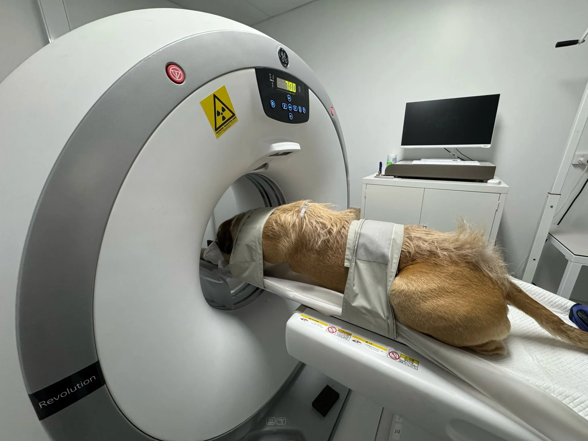
CT
More Information
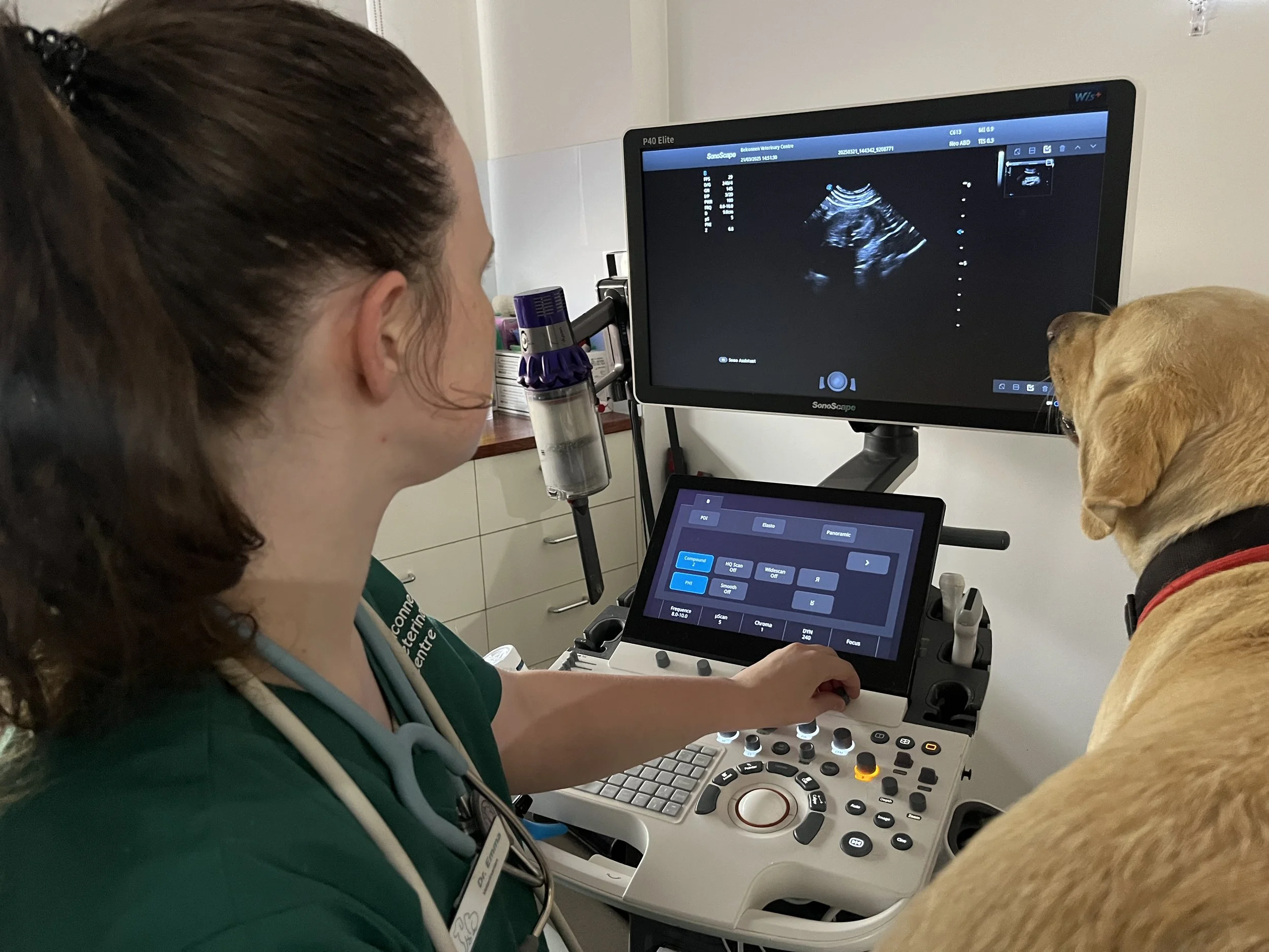
Ultrasound
More Information
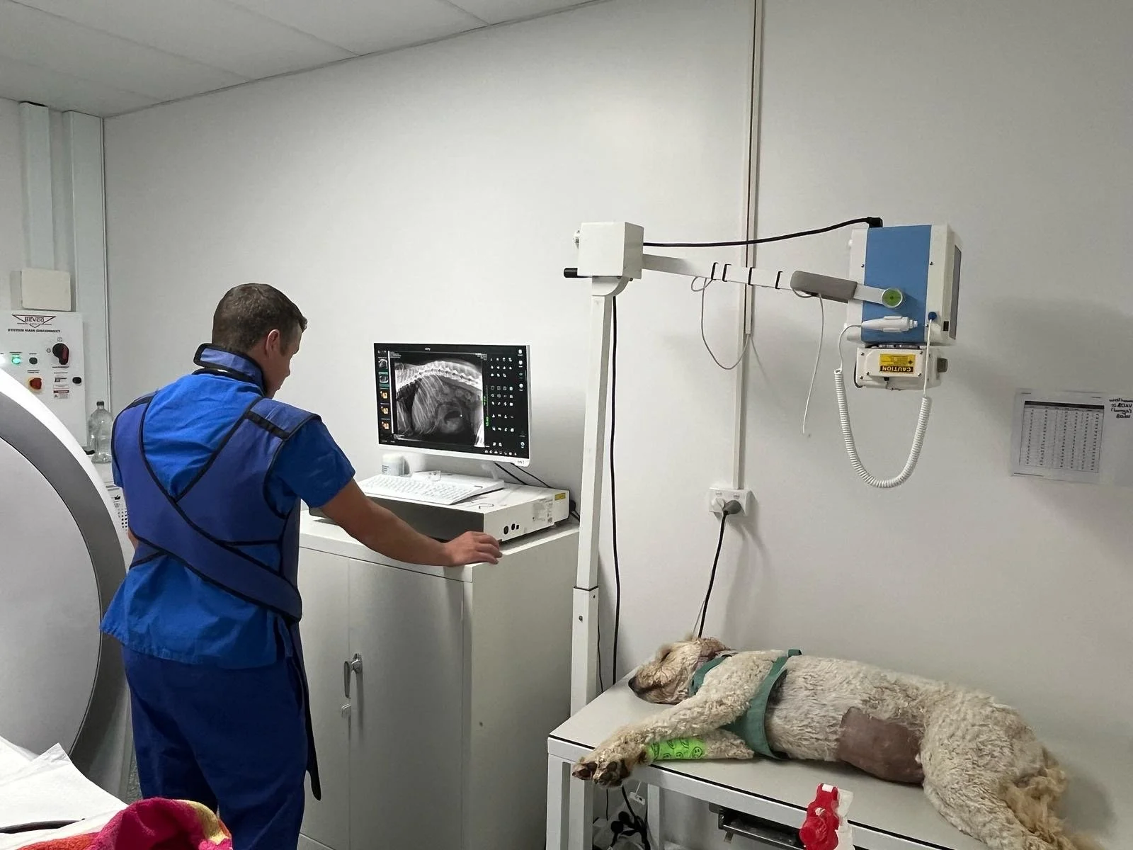
X-Rays
More Information